Services

What is Cerebral Arteriovenous Malformation
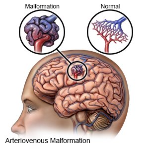
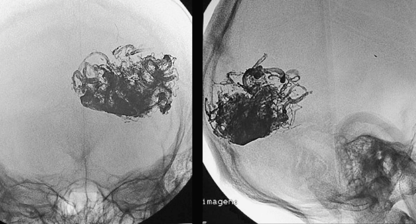
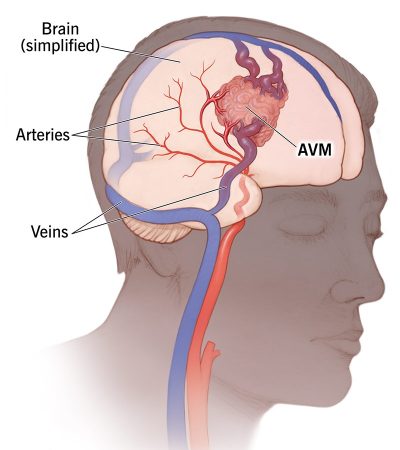
Cerebral arteriovenous malformations (AVMs) is a complex vascular abnormalities characterized by abnormal connections between arteries and veins within the brain or the dura mater, respectively. Dr. Santosh B. Patil offers a comprehensive and multidisciplinary approach to managing these challenging conditions, employing advanced techniques and collaborating closely with neurosurgical experts to ensure optimal patient outcomes.
Symptoms

Symptoms of Cerebral Arteriovenous Malformation
Dural AVF Management
A brain arteriovenous malformation (AVM) may not cause any symptoms until the AVM bursts and bleeds, known as a hemorrhage. In about half of all brain AVMs, a hemorrhage is the first sign of the malformation.

We offers a comprehensive and Multidisciplinary approach to managing these Cerebral Arteriovenous Malformation

Treatment

Endovascular Embolization using Glue/Onyx

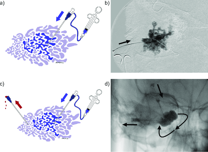
Endovascular embolization is a minimally invasive procedure that involves the selective injection of embolic agents directly into abnormal blood vessels supplying the AVM. Dr. Patil utilizes specialized embolic materials such as glue or Onyx to occlude the abnormal vascular connections and redirect blood flow away from the lesion.
AVM

Glue Embolization

Dr. Santosh Patil carefully navigates microcatheters to the site of the AVM and injects medical-grade glue into the abnormal vessels. The glue solidifies within the vessels, forming a permanent seal and effectively blocking blood flow, thereby promoting the regression of the lesion over time.
AVM

Onyx Embolization

Alternatively, Dr. Santosh Patil may opt for Onyx embolization, a liquid embolic agent with unique properties that enable controlled and precise delivery into the target vessels. Onyx gradually solidifies upon contact with blood, allowing for gradual occlusion of the abnormal vasculature while preserving normal blood flow in adjacent vessels.
AVM

Surgical Excision

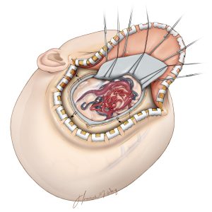
Surgical excision involves the complete removal of the cerebral arteriovenous malformation (AVM) through open surgery. During the procedure, the neurosurgeon carefully dissects around the AVM, isolates the abnormal blood vessels, and removes them from the surrounding brain tissue. Surgical excision is typically recommended for smaller AVMs located in accessible areas of the brain. While effective, surgical excision carries risks such as bleeding, infection, and neurological deficits, and may not be suitable for all patients, especially those with large or deep-seated AVMs.
surgical-excision
AVM

Stereotactic Radiosurgery

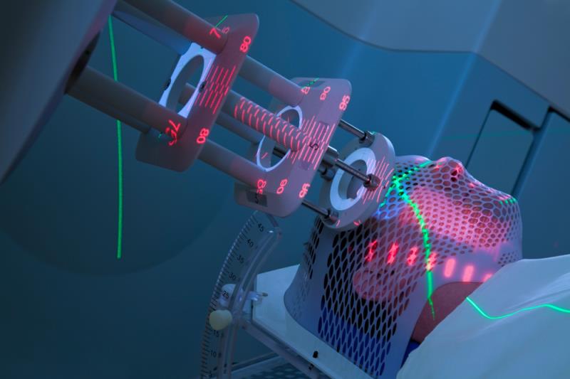
Stereotactic radiosurgery is a non-invasive treatment option for cerebral arteriovenous malformations (AVMs) that utilizes precisely targeted radiation beams to obliterate the abnormal blood vessels. During the procedure, highly focused radiation is delivered to the AVM while minimizing exposure to surrounding healthy brain tissue. Over time, the radiation induces vascular sclerosis and thrombosis within the AVM, leading to its gradual closure. Stereotactic radiosurgery is typically recommended for smaller AVMs or those located in critical or surgically inaccessible areas of the brain. It offers a lower risk alternative to surgical excision, with reduced potential for complications and shorter recovery times.

Dr. Patil collaborates closely with neurosurgical colleagues to perform surgical excision & Stereotactic Radiosurgery (SRS).

Head & Neck Vascular Malformation

FAQ's

Frequently Asked Questions

A cerebral AVM is an abnormal tangle of blood vessels in the brain that disrupts normal blood flow and increases the risk of bleeding or stroke. A dural AVF is an abnormal connection between arteries and veins in the dura mater, the outer covering of the brain.

Symptoms can vary widely depending on the location and size of the lesion but may include seizures, headaches, neurological deficits (such as weakness or numbness), visual disturbances, or symptoms of increased intracranial pressure.

Diagnosis typically involves imaging studies such as magnetic resonance imaging (MRI), magnetic resonance angiography (MRA), or digital subtraction angiography (DSA). These tests help visualize the abnormal blood vessels and assess the extent of the lesion.

Treatment options may include endovascular embolization using glue or onyx, surgical excision, radiosurgery, or a combination of these approaches. The choice of treatment depends on factors such as the size, location, and complexity of the lesion, as well as the patient’s overall health.

Risks may include bleeding, stroke, infection, or damage to surrounding brain tissue. However, the benefits of treatment, such as reducing the risk of hemorrhage or alleviating symptoms, often outweigh the potential risks, especially in symptomatic patients.

The prognosis depends on various factors, including the size and location of the lesion, the effectiveness of treatment, and the presence of any complications. With appropriate treatment and follow-up care, many patients can experience symptom relief and lead fulfilling lives with reduced risk of complications.

We offers a comprehensive and Multidisciplinary approach to managing these Cerebral Arteriovenous Malformation

× How can I help you?